Measuring Protein Function in Biologically Relevant Models
The more biologically relevant your model, the better your predictions. Our solutions help you get more relevant answers from plate-based screens through organoids to whole-animal studies.
Get Biologically Relevant Answers From Plate to Animal
Every assay is a simplification. The question is how much biology you're willing to trade for convenience.
Your protein of interest doesn't live in a tube. It lives in a cell, shaped by its environment in ways no isolated system can replicate. Move into 3D models and you recover the gradients and matrix interactions that govern how compounds penetrate tissue and cells respond. Take it in vivo, and you finally see the pharmacological behavior that determines whether a mechanism becomes a medicine.
We offer solutions built to travel that entire path — sensitive enough to detect proteins at endogenous expression levels, compatible with spheroid and organoid workflows, and bright enough to image in a living animal. The same portfolio of reporters, from a 384-well plate to a whole animal.
Featured Solutions
CRISPR-Edited Cell Models
Cell-Based Assays Using NanoLuc® Technology
3D & Organoid Models
In Vivo Imaging
Frequently Asked Questions
Measuring protein-protein interactions (PPIs) requires choosing between two competing needs: controlled assay conditions or physiologically relevant context. Biochemical assays using purified molecules offer tight experimental control—clean backgrounds, repeatable measurements, and the ability to isolate single interaction events. Live-cell assays capture how PPIs behave in their native context, revealing whether your candidate binds its intended target without triggering unwanted interactions that could compromise safety or efficacy.
When screening compounds at scale, you need the control of a biochemical assay without sacrificing actionable kinetics data. NanoBiT® Technology delivers both: measure PPI binding kinetics in real time using your standard luminescence plate reader. This means you can screen hundreds of compounds while tracking not just whether an interaction occurs, but how fast binding and dissociation happen, which often predicts in vivo behavior.
Measuring target engagement in live cells results in a more accurate analysis of binding between a drug molecule and its target protein. Cell-free assays miss critical real-world factors: off-target binding, cellular crowding, and competition from endogenous proteins. Live-cell measurement captures binding as it actually happens so you identify true binders faster, predict in vivo behavior earlier, and catch liabilities before expensive in vivo studies.
NanoBRET® Technology makes this practical: measure target engagement in standard 96- or 384-well plates without lysis, with ratiometric data that corrects for cell-number variation.
Transient expression is fast and accessible, but it can mask the biology of native-level protein expression. To measure a target protein at its endogenous expression level, CRISPR-based knock-in tagging inserts a small peptide tag directly at the native genomic locus using a short DNA template. This preserves the protein's natural expression context and abundance.
The choice of tag depends on your readout preference. A luminescent tag, such as HiBiT, enables quantitative detection with high sensitivity and low background, and is ideal for high-throughput screening. A fluorescent tag such as HaloTag provides spatial information such as subcellular localization and trafficking, and works well in imaging workflows.
Separating surface-exposed protein from the intracellular pool requires reagents that act selectively at the plasma membrane. The Nano-Glo® HiBiT Extracellular Detection System quantifies only surface-accessible HiBiT-tagged protein in live cells, which is well-suited for tracking receptor internalization. For imaging, Janelia Fluor® HaloTag® Ligands are available in both cell-permeant and impermeant forms, so you can label the surface population alone or the total pool and resolve where your protein sits.
The Nano-Glo® HiBiT Lytic Detection System provides a simple, high-sensitivity endpoint readout, while the extended stability and signal duration of Nano-Glo® Endurazine™ Live Cell Substrate enables real-time kinetic monitoring in intact cells. Pairing either with a NanoBRET® Ternary Complex Kit adds mechanistic confirmation that the degrader is engaging both target and E3 ligase upstream of the degradation readout.
Featured Resources

Imaging
Learn about various imaging techniques, including live cell imaging, in vivo imaging, bioluminescence imaging and fluorescence microscopy.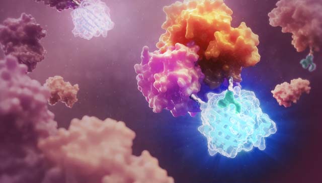
Kinetic Cell-Based Assays
Live-cell kinetic assays allow the same sample well to be repeatedly measured over multiple time points. Get better quantitative data without cumbersome multi-plate setups.