Viral ToxGlo™ Assay
Determine Viral CPE or Monitor Compounds for Antiviral Activity
- Easy, simple protocol measures ATP
- Quantifiable assay for measuring viral-induced cytopathic effect (CPE)
- Compatible with high-throughput screening

Catalog Number:
Size
Catalog Number: G8941
Catalog Number: G8942
Catalog Number: G8943
The Viral ToxGlo™ Assay is a simple, quantifiable method of determining viral-induced cytopathic effects (CPE) in host cells caused by lytic virions. The assay measures cellular ATP as a surrogate measure of host cell viability. When CPE occurs due to viral infection, ATP depletion can be measured and correlated with viral burden. The amount of ATP detected is directly proportional to the number of viable host cells in culture and can be used as a simple method to quantify viral-induced CPE. The homogeneous "add-mix-measure" assay procedure involves adding the single reagent (ATP Detection Reagent) directly to host cells following viral treatment. A “glow-type” luminescent signal is generated that is proportional to the amount of ATP present. Cell washing, multiple pipetting steps and visual assessment are not required to assess CPE. The system detects as few as 15 cells/well in a 384-well format in 10 minutes after reagent addition and mixing and is designed for use in multiwell formats, making it ideal for automated high-throughput screening (HTS).
Features and Benefits
Objectively Quantify CPE: The assay provides quantifiable data by luminescence detection, which obviates subjective operator error associated with visual scoring methods.
Decrease Time to Results: Data can be recorded and analysis begun 10 minutes after reagent addition.
Simplify Assessment of CPE: The homogeneous “add-mix-measure” protocol dramatically reduces the manual steps required for CPE assessment.
Choose Your Format: The reagent is scalable from 96- to 1536-well plate formats.
Amenable to High-Throughput Screening: Luminescent signal is very stable with a half-life generally >5 hours dependent on cell type and medium used, allowing batch or consecutive processing. No fluorescence interference results in high signal to background and delivers excellent Z′ values in screening applications.
Applications include the determination of viral infectivity and the corresponding tissue culture infective dose (TCID50) and potential antiviral potency or off-target toxicity of test compounds.
This method is only useful for viruses that produce cytotoxicity and CPE.
Quantifiable Measure of CPE
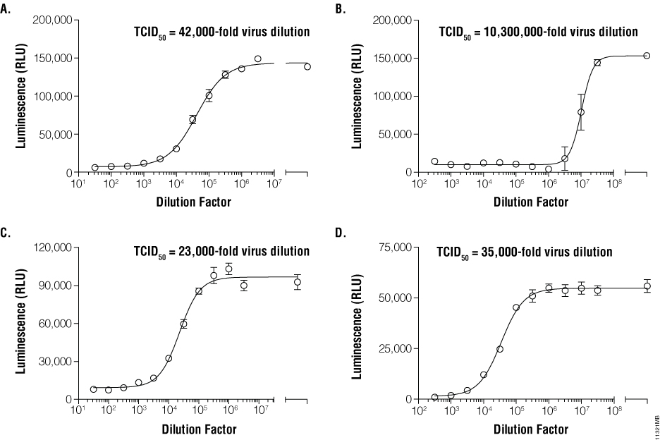
Increased luminescence indicates less infective virus and less CPE. Panel A. Dilutions of Influenza virus H1N1 were applied to MDCK monolayers. Panel B. Dilutions of VEEV applied to Vero E6 monolayers. Panel C. Dilutions of dengue virus applied to BHK-21 monolayers. Panel D. Dilutions of RSV applied to A549 monolayers. After incubation, ATP Detection Reagent was added directly to cells and luminescence measured.
Accurately Calculate Toxicity of Test Compounds
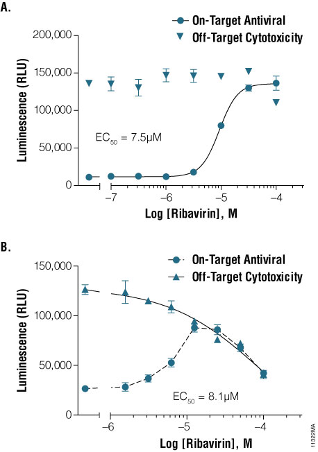
Panel A. Test compound reduces viral CPE with no off-target cytotoxicity. MDCK cells with 100 TCID50 of H1N1 (on-target antiviral) or MDCK cells only (off-target cytotoxicity). Panel B. Test compound reduces viral CPE and also causes off-target cytotoxicity. BHK-21 cell monolayer with 100 TCID50 of Dengue virus (on-target antiviral) or BHK-1 cells only (off-target cytotoxicity). After incubation, ATP Detection Reagent was added directly to cells and luminescence measured.
Protocols
Complete Protocol
Specifications
Catalog Number:
Contenido
| Item | Part # | Presentación |
|---|---|---|
|
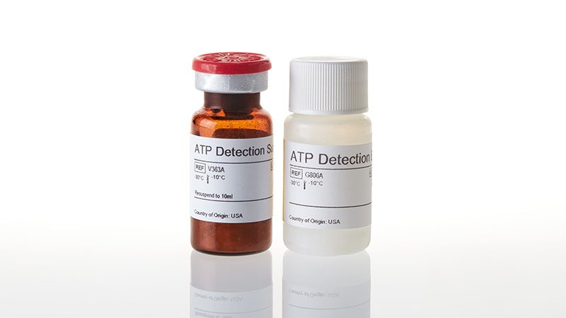
ATP Detection Buffer |
G806A | 1 × 10ml |
|
ATP Detection Substrate |
V363A | 1 × 1 vial |
SDS
Search for SDSCertificado de Análisis
Use Restrictions
For Research Use Only. Not for Use in Diagnostic Procedures.Condiciones de Almacenaje
Contenido
| Item | Part # | Presentación |
|---|---|---|
|
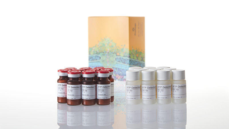
ATP Detection Buffer |
G806A | 10 × 10ml |
|
ATP Detection Substrate |
V363A | 10 × 1 vial |
SDS
Search for SDSCertificado de Análisis
Use Restrictions
For Research Use Only. Not for Use in Diagnostic Procedures.Condiciones de Almacenaje
Contenido
| Item | Part # | Presentación |
|---|---|---|
|
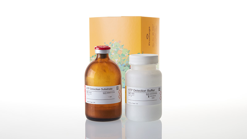
ATP Detection Buffer |
G806B | 1 × 100ml |
|
ATP Detection Substrate |
V363B | 1 × 1 vial |
SDS
Search for SDSCertificado de Análisis
Use Restrictions
For Research Use Only. Not for Use in Diagnostic Procedures.Condiciones de Almacenaje
Resources
Otros
Related Products
Productos Similares
CellTox™ Green Cytotoxicity Assay
Mide los cambios en la integridad de la membrana. Monitoriza cinéticamente la citotoxicidad hasta 72 horas y además tiene capacidad multiplex.
G8741, G8742, G8743, G8731
MultiTox-Glo Multiplex Cytotoxicity Assay
Ensayo fluorescente y luminiscente que le permite medir el número relativo de células vivas y muertas en una población.
G9270, G9271, G9272
CytoTox-Glo™ Cytotoxicity Assay
Ensayo de citotoxicidad basado en luminiscencia, de alta sensibilidad que mide el número relativo de células muertas.
G9290, G9291, G9292
Maxwell® RSC Viral Total Nucleic Acid Purification Kit
Extracción de ácidos nucleicos totales virales a partir de varios tipos de muestras, utilizando el Maxwell® RSC Instrument.
AS1330, ASB1330
Usado con frecuencia junto con
BacTiter-Glo™ Microbial Cell Viability Assay
Mida el número de células microbianas viables en cultivo mediante una señal luminiscente proporcional a la cantidad de ATP presente.
G8230, G8231, G8232, G8233
CellTiter-Blue® Cell Viability Assay
Ensayo fluorescente, homogéneo y con resazurina que permite monitorizar la viabilidad celular.
G8080, G8081, G8082
CellTiter-Fluor™ Cell Viability Assay
Ensayo de viabilidad celular fluorescente no lítico con capacidad multiplex.
G6080, G6081, G6082
GloMax® Discover System
Lector de microplacas de alto rendimiento para la detección de luminiscencia, fluorescencia y absorbancia.
GM3000