Lumit® Insulin Immunoassay
No-Wash Immunoassay for Insulin Detection
- Fast add-and-read protocol
- Luminescent detection with just a plate-reading luminometer
- Flexible, scalable throughput: Perform 100 assays in 96-well format or 400 assays in 384-well format using W8010; perform 500–2000 assays with W8012
Catalog Number:
Size
Catalog Number: W8010
Catalog Number: W8012
Detect Insulin Secretion Without Tedious Wash Steps
The Lumit® Insulin Immunoassay quantitatively measures insulin from cell culture or islet secretion samples with a fast, easy, no-wash protocol. Simply add Lumit® reagents to the sample, incubate and read using a standard multimode plate reader. Results are available in just 70 minutes. Lumit® assays are ideal for quantitating hormone secretion from islet perifusion workflows. The flexible assay format can be scaled to 384-well plates for high-throughput sample processing.
How the Lumit® Insulin Immunoassay Works
Primary antibodies to insulin, selected for their specificity and sensitivity, are labeled with the LgBiT and SmBiT subunits of NanoBiT® Luciferase. In the presence of insulin, the subunits are brought together to reconstitute active luciferase enzyme. Adding the optimized luciferase substrate generates a bright luminescent signal that is proportional to analyte levels.

Simple One-Plate Protocol, No Wash Steps

Recommended Multiwell Assay Formats
|
Plate Format |
Insulin Sample |
Total Reaction |
Number of Assays |
Number of Assays |
|
96-Well |
50µl |
125µl |
100 |
500 |
|
384-Well |
12.5µl |
31.1µl |
400 |
2000 |
Lumit® assay setup is highly flexible and can be scaled up or down as desired. In addition to the configurations listed above, the assay is compatible with any sample volume ≥5µl provided proportionate amounts of Lumit® reagents are used. For more information, see Table 1 in the Lumit® Insulin Immunoassay Technical Manual (#TM776).
Lumit® Insulin Immunoassay Has Broad Range and Picomolar Sensitivity
The picomolar to nanomolar linear range accommodates popular cell models and islets.


|
Specification |
Lumit® Insulin Immunoassay |
|
|
Limit of Detection (LOD) |
10pM |
|
|
Range |
10pM–8nM |
|
|
Signal/Background max |
>200 |
|
|
Assay Time |
70 minutes |
|
|
Sample Type |
Cell culture supernatants |
|
SD=Standard Deviation
Note: Lumit® Insulin Immunoassay kits do not include a positive control. Instructions for preparing your own positive control for use with the assay can be found in TM776.
Applications for the Lumit® Immunoassays
Insulin and glucagon are key target analytes measured in metabolism research. Secreted from β and α cells of pancreatic islets, respectively, the two hormones are vital for regulating glucose levels in the body. Using the Lumit® Insulin and Lumit® Glucagon Immunoassays, it is easy to collect hormone secretion data from monolayer cells in culture, 3D islet microtissues and islets. The simple add-and-read protocol is compatible with static and dynamic glucose-stimulated insulin secretion (GSIS) experiments, including perifusion systems.
Perifusion is a powerful method for studying hormone secretion over time and in response to sequential treatments, and it generates a large number of samples for analysis. Lumit® Immunoassays make analysis of large sample sets like this much easier by avoiding tedious wash steps. The faster time to result gives you data sooner and enables you to move on to your next experiment.
Quantitate Insulin and Glucagon Secretion from Mouse Pancreatic Islets

Insulin and glucagon secretion were measured in samples collected during perifusion experiments. Briefly, 80 mouse islets were placed in triplicate chambers of a perifusion instrument (Biorep, Miami Lakes, Florida). The islets were treated with 2.7mM glucose and then 10mM glucose, in combination with an amino acid mixture. Perifusate was collected every minute. Ten microliters of each sample were transferred into wells of a 384-well plate and assayed for either insulin or glucagon. Insulin and glucagon data sets are superimposed. This data was kindly provided by Drs. Hannah Foster and Matthew Merrins, University of Wisconsin VA Hospital, Madison, Wisconsin.
Featured Application Note
Measuring GLP-1 Receptor Agonist Activity in Cells with the Lumit® Insulin Immunoassay
Imperial College London used the Lumit® Insulin Immunoassay to differentiate the potency and activity of three GLP-1 receptor agonist drug candidates, demonstrating that the assay is well-suited for high-throughput metabolic disease drug discovery workflows.

Cells were treated with three GLP-1 receptor agonist drug candidates and insulin secretion measured using the Lumit® Insulin Immunoassay. Each compound demonstrated distinct potency and maximal response, highlighting the assay's ability to discriminate pharmacological differences across candidates.
Protocols
Complete Protocol
Specifications
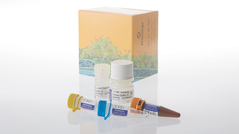
Catalog Number:
Contenido
| Item | Part # | Presentación |
|---|---|---|
Lumit® Detection Substrate B |
VB405A | 1 × 160μl |
Lumit® Detection Buffer B |
VB406A | 1 × 3.2ml |
Lumit® Antibody Dilution Buffer B |
W150A | 1 × 5.5ml |
Lumit® Anti-Insulin mAb-SmBiT, 200X |
W152A | 1 × 30μl |
Lumit® Anti-Insulin mAb-LgBiT, 200X |
W153A | 1 × 30μl |
SDS
Search for SDSCertificado de Análisis
Use Restrictions
For Research Use Only. Not for Use in Diagnostic Procedures.Condiciones de Almacenaje
U.S. Pat. No. 8,809,529, European Pat. No. 2635582, Japanese Pat. No. 5889910 and other patents and patents pending.
U.S. Pat. Nos. 9,797,889, 9,797,890, 10,107,800 and 11,493,504; European Pat. No. 2970412; Japanese Pat. Nos. 7280842 and 7532562; and other patents and patents pending.
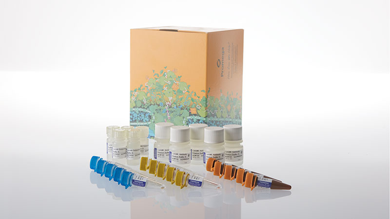
Contenido
| Item | Part # | Presentación |
|---|---|---|
Lumit® Detection Substrate B |
VB405A | 5 × 160μl |
Lumit® Detection Buffer B |
VB406A | 5 × 3.2ml |
Lumit® Antibody Dilution Buffer B |
W150A | 5 × 5.5ml |
Lumit® Anti-Insulin mAb-SmBiT, 200X |
W152A | 5 × 30μl |
Lumit® Anti-Insulin mAb-LgBiT, 200X |
W153A | 5 × 30μl |
SDS
Search for SDSCertificado de Análisis
Use Restrictions
For Research Use Only. Not for Use in Diagnostic Procedures.Condiciones de Almacenaje
U.S. Pat. No. 8,809,529, European Pat. No. 2635582, Japanese Pat. No. 5889910 and other patents and patents pending.
U.S. Pat. Nos. 9,797,889, 9,797,890, 10,107,800 and 11,493,504; European Pat. No. 2970412; Japanese Pat. Nos. 7280842 and 7532562; and other patents and patents pending.
Resources
Featured Citation
In this work, El and colleagues use the Lumit® Glucagon Immunoassay to characterize the complex regulatory mechanisms that control glucagon secretion from alpha cells.
El, K. et al. (2021) GIP mediates the incretin effect and glucose tolerance by dual actions on α cells and β cells. Sci. Adv. 7, abf1948.
Artículos
- Understanding the Complicated Relationship Between Insulin, Glucagon and GIP
- Blog Article: Jon Campbell Is Challenging Classic Models of Metabolic Disease
- Blog Article: Infographic: Assays for Measuring Insulin Activity
- White Paper: GLP-1 Signaling in Obesity Technologies for Monitoring GPCR Dynamics
- Application Note: Measuring Insulin Release Following GLP-1 Receptor Agonist Treatment using the Lumit® Insulin Immunoassay
Posters
Related Products
Productos Similares
Lumit® Glucagon Immunoassay
Mide el glucagón de forma cuantitativa a partir de muestras de cultivos celulares o de secreción de islotes mediante un protocolo rápido y sencillo que no requiere lavado.
W8020, W8022
Lumit® IL-1β Human/Mouse Immunoassay
Cuantifica la activación del inflamasoma mediante la medición de la IL-1β liberada utilizando un protocolo sencillo y sin lavado.
W6010, W6011, W6012, W7010, W7011, W7012, W116A-C, W119A-C
Lumit® TNF-α (Human) Immunoassay
Mide cuantitativamente el TNF-α liberado en muestras de cultivos celulares mediante un protocolo sencillo y sin lavado.
W6050, W6052, W6051, W137A-C
Lumit® IL-6 (Human) Immunoassay
Mide cuantitativamente la IL-6 liberada en muestras de cultivos celulares mediante un protocolo sencillo y sin lavado.
W6030, W6032, W6031, W128A-C
Usado con frecuencia junto con
Glucose Uptake-Glo™ Assay
Ensayo no radiactivo para medir la captación de glucosa.
J1341, J1342, J1343
Triglyceride-Glo™ Assay
Detecta los niveles de triglicéridos midiendo el glicerol que se libera de una reacción enzimática con una lipasa.
J3160, J3161
Glycogen-Glo™ Assay
Método rápido y sensible para la detección de glucógeno en muestras biológicas.
J5051, J5052
Incretin-Related Bioassays
Metabolic disease bioassays designed for potency testing and functional analysis.
CS3629B22, CS3629B24, CS3629B26, CS3629B15, CS3629B17, CS3629B19, CS3629B05, CS3629B07, CS3629B02, CS354805, CS354807, CS354802