How to Get Started with Multiplexing Cell-Based Assays
Learn how multiplexing cell-based assays lets you collect multiple readouts from the same sample, such as viability, cytotoxicity, and metabolic data, and get practical tips on assay compatibility and proven workflows for 2D cultures and 3D models like organoids and spheroids.
Simon Moe, Promega Corporation
Publication date: November 2026
What is multiplexing?
Multiplexing refers to getting more than one set of data from the same sample. In the context of cell-based assays, this is typically done by running multiple assays from same sample of wells, as opposed to using each well for 1 single readout. If a standard workflow using an endpoint viability assay is paired with a real-time cytotoxicity assay, the amount of data you get per well is vastly increased.
Why multiplex?
Consider the following. You ran a large screen using a viability assay and narrowed down to a few candidates. Months later you realize the treatments weren’t killing the cells, they were just cytostatic. Those 3 months and thousands of dollars were wasted because you didn’t check cell death right away. This has been shown with organoid models of pancreatic cancer, where multiplexing CellTiter-Glo® 3D Cell Viability Assay and CellTox™ Green Cytotoxicity Assay showed that after treatment with anticancer agents, some cells showed a reduction in viability, indicated by a drop in ATP, but did not have a simultaneous increase in cell death (Mäkinen et al., 2022).
One of the most straightforward reasons to multiplex is to ensure you have a ‘control’ for your in vitro experiments. A good example of this would be to include a viability marker such as CellTiter-Fluor™ Cell Viability Assay alongside a reporter assay to ensure that any change in expression is real and not simply due to cells dying. A separate approach would be to multiplex several biologically related readouts from one sample, to gain a richer understanding of a pathway. An example of this would be multiplexing several assays that detect individual metabolites to more fully characterize metabolic signaling.
Related ResourcesLearn more in this blog: Save Precious Time with Same-Well Multiplexing - Promega Connections
Practical Tips for Multiplexing Cell-Based Assays
There are several important considerations when multiplexing cell based assays. These include the following:
- Assay volumes must fit in the volume of the assay well or be easily physically separated.
- Signal readouts from each assay must be spectrally distinct.
- Assay chemistries must be compatible (e.g., avoid components that might precipitate or interfere with assay signal).
- Assays using nonlytic reagents should be performed before introducing assays with cell lysis reagents. This may seem obvious, but reviewing the principle behind the assay chemistries will help map out your multiplex procedure.
- You may need to use a more concentrated assay working reagent for the first assay to leave enough room in the well to add the appropriate volume of the second assay reagent. Consult the assay manufacturer’s technical information for any instructions to concentrate their assay reagent.
- White, opaque plates provide higher signals for fluorescent or luminescent readouts and may reduce the upper limit of the multiplex assay exhibiting saturation. However, sensitivity may not increase since there will be higher backgrounds as well. Black, opaque plates have lower signal and background responses, but very little cross-talk or light scattering compared to white, opaque plates. If a luminescent signal varies more than 2 logs between replicates, then use black, opaque plates to limit crosstalk.
- When combining signal readouts, it is not practical to perform colorimetric assays in the same well as fluorescent or luminescent assays due to the ability of the chromogen to quench these signals. If the sample can be physically divided or separated (culture medium vs cells) into different wells, then the assay chemistries will not interfere with each other.
- Combining multiple fluorescent assays should be possible so long as the following is ensured:
- The readouts are spectrally distinct.
- There is no resonance transfer.
- There are no quenching agents (e.g., detergents, redox) affecting companion fluorophores.
- There is minimal autofluorescence.
- Promega has had great success in combining cell-based assays with fluorescent and luminescent readouts. The multiplex assay format typically begins with fluorescent assays (nonlytic) followed by luminogenic assays (lytic). An example of this format is the ApoTox-Glo™ Triplex Assay where the detection of multiple biomarkers occurs sequentially.
Additional information in interpreting multiplexing data can be found here.
Proven combinations: 2D workflows
There are several common approaches to multiplexing in 2D models. The first, and simplest (workflow A) is through the pairing of two assays that have distinct outputs, such as fluorescence and luminescence assays. Another approach (workflow B) aliquots samples and runs separate assays on those distinct samples. This is typically done with assays that use similar chemistry/signal output, such as two luminescence assays that both read biology from the sample medium. Even if assays share bioluminescence as a readout, running them separately from media fractions is still meaningful multiplexing. You can also multiplex two bioluminescent assays if your second assay utilizes lytic components that destroy the first assays enzymes. Below we highlight two brief workflows for these two approaches.
Workflow A: Same-well multiplex:
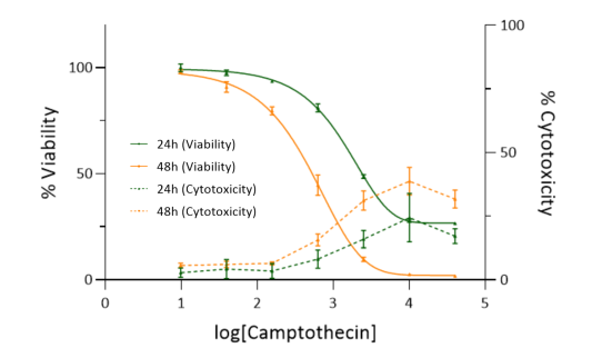
Here we highlight automated multiplexing of RealTime-Glo™ MT Cell Viability Assay and CellTox™ Green Cytotoxicity Assay on the CyBio FeliX.
- Plated U2-OS cells (human osteosarcoma epithelial adherent cells) (initial density of 5,000 cells/well) with RealTime-Glo™ and CellTox™ Green reagents.
- A 4-fold, 7-step serial dilution of Camptothecin (a DNA topoisomerase I inhibitor, inducing apoptosis in a dose-dependent manner) was used and applied. Luminescence and fluorescence were recorded at 24 and 48 hours after treatment.
- Percent viability was calculated and plotted for each assay against the log10[Camptothecin] using a five-parameter logistic equation on the left y axis.
- Percent cytotoxicity was calculated and plotted for each assay against the log10[Camptothecin] without regression (mean and connecting lines) on the right y axis.
Read the full protocol in the Application Note.
Workflow B: Sequential/pseudo-multiplex:
- Culture media was removed from individual 3D brain organoids (A, B, and C) on Day 3 (Embryoid Body stage) and Day 30 (Cerebral Organoid stage).
- Glutamate and BCAA concentrations were measured using the Glutamate-Glo™ and BCAA-Glo™ Assays using separate aliquots of sample media.
- Tracking these metabolites during neurodifferentiation can help researchers assess neuronal maturation, synaptic function, and metabolic adaptations associated with brain development.
Read the full protocol in the Application Note.

Multiplexing Assay Solutions
Promega offers a wide range of multiplexable assay solutions, as shown below.

Multiplexing in 3D models
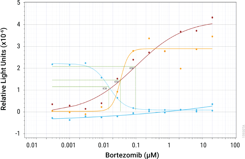
3D model systems include organoids, spheroids, and organs on chips. These samples provide a clear example where the time and money saved through multiplexing is most valuable. 3D samples can take months to generate, and often have high sample-to-sample differences due to the more complex nature of the tissues. Here is where multiplexing shines, allowing one 3D system to have multiple parameters measured. Below is an example of multiplexing cytotoxicity and viability from a 3D system.
- Spheroid human colorectal carcinoma microtissues (HTC116) were grown in Ultra-Low Attachment Plates for 3 days at 37°C, 5% CO2 and subsequently treated with Bortezomib (37°C, 5% CO2) to induce LDH release.
- Media samples were taken from the spheroid plate at 24hrs, 48hrs and 64hrs and diluted with LDH Storage Buffer [200mM Tris-HCl (pH 7.3), 10% Glycerol, 1% BSA].
- The diluted samples were assayed with the LDH-Glo™ Cytotoxicity Assay.
- Spheroids were then assayed with the CellTiter-Glo® 3D Cell Viability Assay.
- The LDH curves are represented with circles: blue at 24hrs, red at 48hrs and yellow at 64hrs. The CellTiter-Glo® curve is represented with blue diamonds and a dotted line as a single endpoint curve.
Read the full protocol in the Application Note.
Related Resources
Interested in more solutions for multiplexing from 3D models? Explore the full selection of tools to monitor 3D cell biology.
Want to see another example of multiplexing using a 3D cell culture? Read more to see how LDH-Glo™, CellTiter-Glo®, and P450-Glo® assays were used to assess toxicology on stem cell-derived OrganoReady Colon Organoids (Mimetas & Promega, 2025).
Citations
- Hsieh, J. H., Huang, R., Lin, J. A., Sedykh, A., Zhao, J., Tice, R. R., Paules, R. S., Xia, M., & Auerbach, S. S. (2017). Real-time cell toxicity profiling of Tox21 10K compounds reveals cytotoxicity dependent toxicity pathway linkage. PLoS ONE, 12(5), Article e0177902. https://doi.org/10.1371/journal.pone.0177902
- Klatt, S., Marquardt, G., Faxel, M., Rubner, S., & Papasotiriou, I. (2026). Comparison of THP-1 Subtypes Reveals Differences on Transcriptomic and Protein Expression Level Throughout Their Immunological Differentiation. Journal of Immunology Research, 2026(1), Article 9427482. https://doi.org/10.1155/jimr/9427482
- Mäkinen, L., Vähä-Koskela, M., Juusola, M., Mustonen, H., Wennerberg, K., Hagström, J., Puolakkainen, P., & Seppänen, H. (2022). Pancreatic Cancer Organoids in the Field of Precision Medicine: A Review of Literature and Experience on Drug Sensitivity Testing with Multiple Readouts and Synergy Scoring. Cancers, 14(3), Article 525. https://doi.org/10.3390/cancers14030525
- Mimetas & Promega. (2025). Predicting Gastrointestinal Toxicity using Adult Stem Cell-derived OrganoReady Colon Organoid with Functional Multiplexed Assays. [Application note].