LDH-Glo™ Cytotoxicity Assay
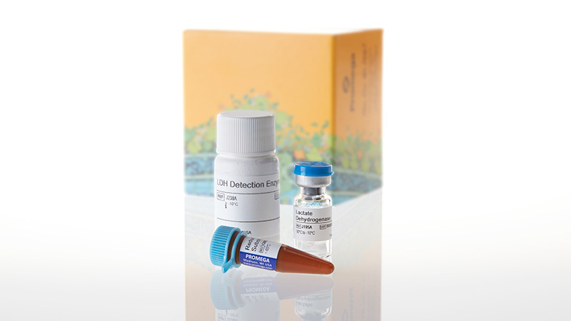
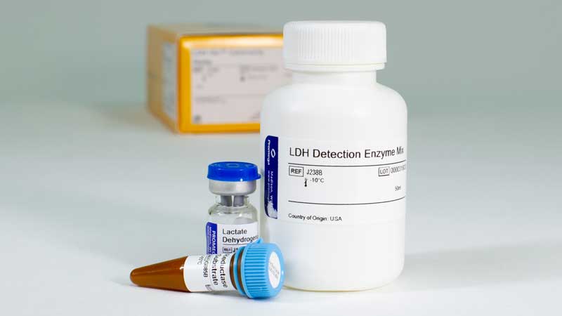
Simple, More Sensitive LDH Assay Method to Determine Cytotoxicity
- Detects LDH release from small numbers of cells, including 3D cell models
- Monitors cytotoxicity from the same sample over time
- Provides more data per well through multiplexing with other cell-based assays
- Integrates seamlessly with the MyGlo® Reagent Reader

Catalog Number:
Choose a product
Size
Catalog Number: J2380
Catalog Number: J2381
Monitor Cytotoxicity from Small Numbers of Cells
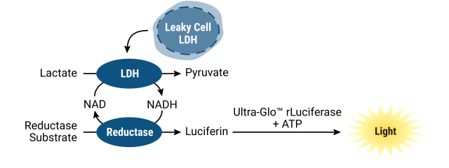
The LDH-Glo™ Cytotoxicity Assay is a bioluminescent plate-based assay for quantifying lactate dehydrogenase (LDH) release into the culture medium upon plasma membrane damage. The bioluminescent detection is more sensitive than colorimetric or fluorescent methods, allowing accurate detection of LDH from a small number of cells, including primary cells and 3D cell cultures.
This LDH assay involves removing only a small amount of cell media (2–5µl) from each treated well, allowing you to get more data by sampling the same well over time and by using the remaining media and cells for other cell-based assays.
How Does the LDH-Glo™ Cytotoxicity Assay Work?
How Do I Monitor Cytotoxicity in 3D Cell Cultures?
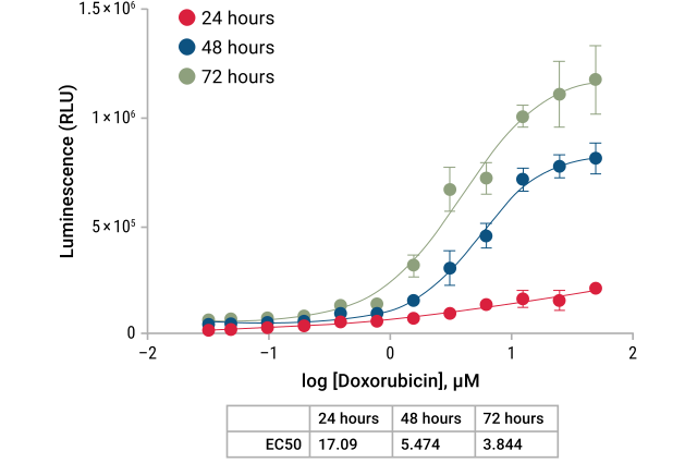
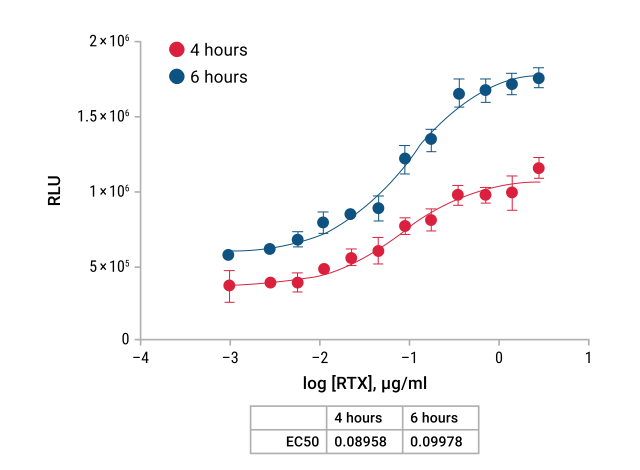
The LDH-Glo™ Cytotoxicity Assay is well suited for measuring LDH released from small numbers of membrane-damaged cells. Here is an example of measuring drug-induced toxicity in HCT116 spheroids. The time-dependent toxicity measurements were performed by repeatedly sampling media from the same wells.
How do I run the LDH-Glo™ assay with 3D spheroids or organoids, and what controls should I include?
Remove just 2–5µl of medium per timepoint—up to about 10% of a well's starting volume doesn't measurably affect remaining cells or spheroids—dilute into LDH Storage Buffer, and freeze or assay directly. Every plate should include a No-Cell Control (medium background), a Vehicle-Only Control, and, if calculating percent cytotoxicity, a Maximum LDH Release Control using Triton™ X-100.
Because 3D models can have heterogeneous baseline LDH release, normalize to a same-plate, same-treatment-day control rather than comparing raw luminescence values across separate experiments.
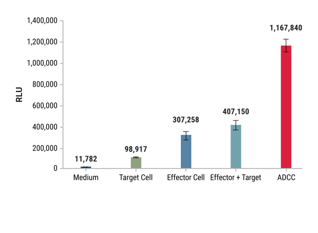
How Do I Detect Target Cell Cytotoxicity in ADCC Assays?
Pair with the MyGlo® Reagent Reader
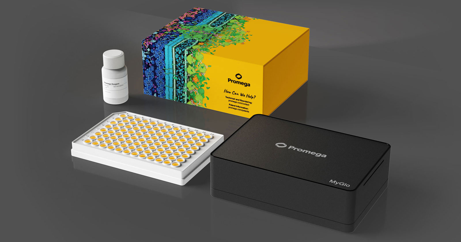
Reading your assay results doesn't require a shared plate reader. The MyGlo® Reagent Reader is a compact 96-well luminescence plate reader, enabling you to run luminescent assays at your bench without competing for instrument time. Integrated software generates dose response curves, single concentration and linearity analysis in minutes.
Protocols
Complete Protocol
Frequently Asked Questions
How can the LDH-Glo™ assay use only 2–5µl of medium for repeated sampling?
Because bioluminescent detection is highly sensitive, only a very small sample is needed to generate a measurable signal. Removing 2–5µl—up to roughly 10% of a well's starting medium volume across several timepoints—doesn't measurably affect the remaining cells, so the same well can be sampled repeatedly to build a time-course without disturbing the culture.
What controls are recommended for LDH-Glo™ assay in 3D cultures?
Every assay plate should include a No-Cell Control (triplicate wells without cells, to measure medium background) and a Vehicle-Only Cells Control (untreated cells with the same solvent used for test compounds). If you need percent cytotoxicity, add a Maximum LDH Release Control by treating vehicle-only cells with Triton™ X-100 to lyse them completely before sampling.
Can LDH-Glo™ assay samples be frozen and measured later?
Yes. Samples frozen in LDH Storage Buffer show no significant loss of LDH activity after a freeze-thaw cycle, but samples frozen directly in PBS with BSA can lose up to 85% of LDH activity—so always store in LDH Storage Buffer before freezing, not PBS.
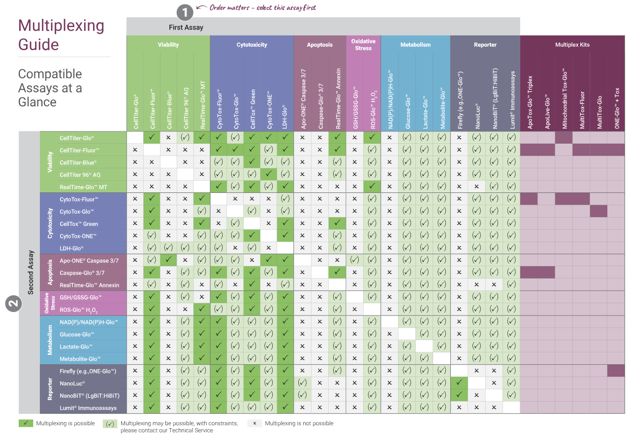
Can the LDH-Glo™ assay be multiplexed with a cell viability assay?
Yes. Because only a small volume of medium is removed for LDH-Glo™ detection, the remaining sample can be used for RealTime-Glo™ MT, CellTiter-Glo® or CellTiter-Fluor™ viability measurements, or for orthogonal endpoints like Caspase-Glo® 3/7, Glucose-Glo™ or Glutamate-Glo™ assays—helping distinguish cytostatic from cytotoxic effects in the same well.
How do you choose the right sample dilution factor for the LDH-Glo™ assay?
Dilution depends on cell number and whether serum is present, since serum itself contains LDH. Promega's technical manual recommends roughly 5-fold dilution for under 1,000 cells/100µl without serum, up to 100-fold for 10,000–50,000 cells/100µl without serum, and up to 300-fold with 10% serum at higher cell numbers—always confirm results fall within the assay's linear range.
Why is bioluminescent LDH detection more sensitive at low signal levels than colorimetric detection?
Bioluminescence produces light against an essentially dark background, so even small amounts of signal are easy to distinguish from noise. Colorimetric detection instead measures a small color change against a comparatively bright background (transmitted light), which requires more analyte to produce a reliably measurable difference—making it less suited to low-cell-number or low-LDH samples.
Specifications
Catalog Number:
What's in the box?
| Item | Part # | Size |
|---|---|---|
Reductase Substrate |
G885A | 1 × 55μl |
Lactate Dehydrogenase |
J195A | 1 × 1 each |
LDH Detection Enzyme Mix |
J238A | 1 × 10ml |
SDS
Search for SDSCertificate of Analysis
Use Restrictions
For Research Use Only. Not for Use in Diagnostic Procedures.Storage Conditions
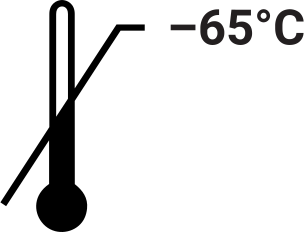
U.S. Pat. Nos. 9,273,343 and 9,951,372, European Pat. No. 2751089, Japanese Pat. No. 6067019 and other patents pending.
What's in the box?
| Item | Part # | Size |
|---|---|---|
Reductase Substrate |
G885B | 1 × 275μl |
Lactate Dehydrogenase |
J195A | 1 × 1 each |
LDH Detection Enzyme Mix |
J238B | 1 × 50ml |
SDS
Search for SDSCertificate of Analysis
Use Restrictions
For Research Use Only. Not for Use in Diagnostic Procedures.Storage Conditions
U.S. Pat. Nos. 9,273,343 and 9,951,372, European Pat. No. 2751089, Japanese Pat. No. 6067019 and other patents pending.
Related Products
Similar Products
CellTox™ Green Cytotoxicity Assay
Measures changes in membrane integrity. Kinetically monitors cytotoxicity up to 72 hours with multiplex capability.
G8741, G8742, G8743, G8731
CytoTox-Glo™ Cytotoxicity Assay
Highly sensitive luminescent cytotoxicity assay that measures the relative number of dead cells.
G9290, G9291, G9292
CytoTox-ONE™ Homogeneous Membrane Integrity Assay
A homogeneous, LDH-based fluorometric method for quantifying non-viable cells.
G7890, G7891, G7892
CytoTox-Fluor™ Cytotoxicity Assay
A single-reagent-addition, homogeneous, fluorescent assay that measures the relative number of dead cells in cell populations.
G9260, G9261, G9262
Frequently Used With
CellTiter-Glo® 3D Cell Viability Assay
A homogeneous method optimized to assess viability in 3D cell culture.
G9681, G9682, G9683
GloMax® Discover System
High-performance microplate reader for detecting luminescence, fluorescence and absorbance.
GM3000
RealTime-Glo™ MT Cell Viability Assay
A bioluminescent method to kinetically monitor viability in cell culture up to 72 hours.
G9711, G9712, G9713
Caspase-Glo® 3/7 Assay System
An easy-to-use, plate-based luminescent assay for detecting caspase-3/7 activity.
G8090, G8091, G8093, G8092
Resources
Articles
- "Will This Drug Cause Hepatotoxicity?"
- App Note: Predicting gastrointestinal toxicity
- Impact of aerosols on liver xenobiotic metabolism: A comparison of two methods of exposure
- Folate and Vitamin B12 Deficiency Exacerbate Inflammation during Mycobacterium avium paratuberculosis (MAP) Infection.
- SARS‐CoV‐2‐reactive T‐cell receptors isolated from convalescent COVID‐19 patients confer potent T‐cell effector function